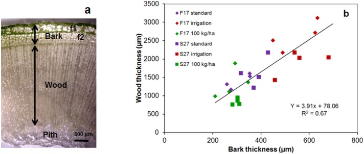
Fig 1. Stereomicroscopy imaging of transverse cross section of the hemp stem.
(a) showing wood and bark including primary fibers (f1), secondary fibers (f2); arrows indicate the dimensions measured to determine wood-/bark-thickness (b) in the basal stem region of F17 and S27 grown under different conditions (standard, irrigation, high sowing density: 100 kg.ha-1).

