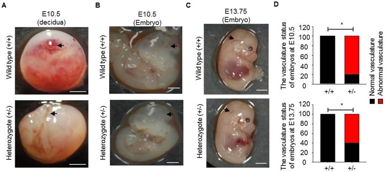
Fig 5. The aberrant vasculature formation is observed in Runx1t1 deficient mouse embryos.
(A) Images showing the vascularity of deciduas from a wild-type and a heterozygous Runx1t1 knockout mouse at embryonic day 10.5 (E10.5). Scale bar = 1mm. Arrows indicated vessels formed. (B-C) Representative images showing vascularity of embryos from wild-type and heterozygous Runx1t1 knockout mice at embryonic day 10.5 (B) and 13.75 (E13.75) (C) Scale bar = 1 mm. Arrows indicated cerebral vessel (CV) formed. (D) The histogram for showing percentage of aberrant vasculature in Runx1t1 deficient embryos at E10.5 (upper panel) and E13.75 (lower panel). n (total mice number used for analysis) = 4, 5, 7, 5 for wild-type embryos (E10.5), Runx1t1 deficient embryos (E10.5), wild-type embryos (E13.75) and Runx1t1 deficient embryos (E13.75), respectively. *, p<0.05, (Fisher’s exact test).

