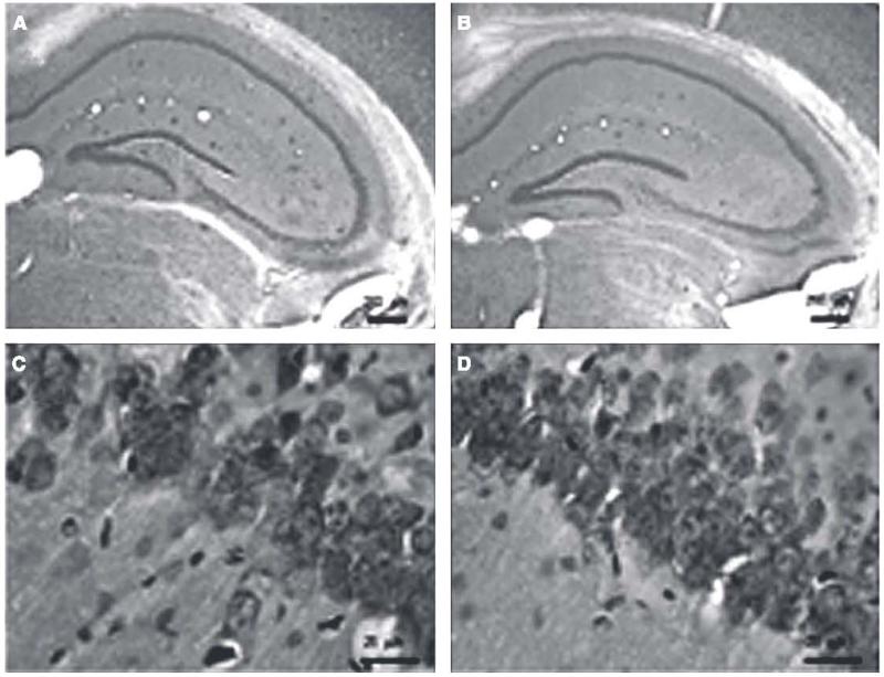
Fig. 3.
Low-magnification micrograph images of Congo red staining of Aβ plaques in the hippocampus region of dtg AβPPswe/PS1ΔE9 mice treated with either placebo (A) or 17αE2 (B). High-magnification images of pyramidal neurons stained with violet in the CA1 region of the hippocampus in placebo-treated (C) and 17αE2-treated (D) dtg AβPPswe/PS1ΔE9 mice.

