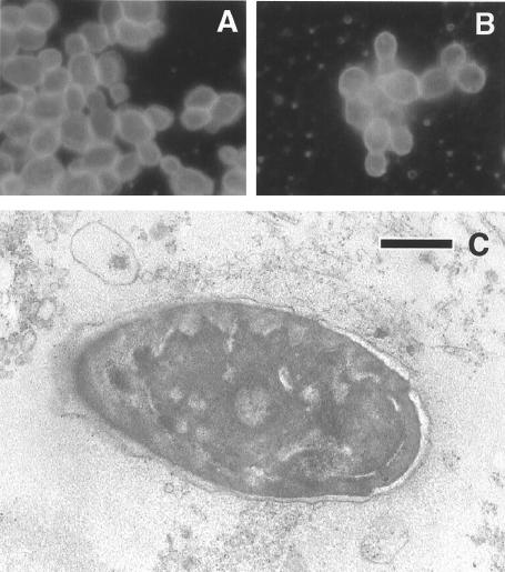
FIG. 1.
Morphological analyses of yeast cells. Comparison of C. famata from the conjunctival exudate (A) with the type culture isolate (B) by immunofluorescence analysis. Immunofluorescence was carried out as described in Materials and Methods. (C) Electron microscopy of yeast cells present in the conjunctival exudates in September 1999. The exudate was placed in PBS and centrifuged. The pellet was processed for electron microscopy. Bar, 200 nm.

