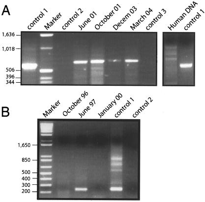
FIG. 2.
PCR assays. (A) PCR of different conjunctival exudate samples. DNA extracted from the exudates was analyzed by PCR, as indicated in Materials and Methods. (B) PCR analysis of different blood serum samples. A total of 200 μl of blood serum obtained from the patient on the dates indicated above the lanes was used for the extraction of total DNA. PCR assays were carried out with oligonucleotides that amplify a region of the rRNA internal transcribed spacer genes. Control 1, positive control consisting of DNA extracted from C. famata; control 2, negative PCR control without DNA; control 3, negative control of DNA extraction consisting of phosphate buffer. The numbers to the left of each gel are in base units.

