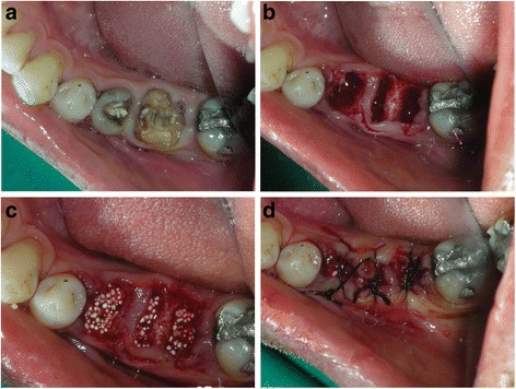
Fig. 1.

a Clinical occlusal view with fractured 45 and 46. b Post-extraction view of the socket. Note minimal trauma to the soft tissue and no flap reflection on the surgical site. c Graft material condensed into the extraction sockets showing good initial graft stability. d Black silk sutures placed with tissue approximation and no releasing incision in the flaps
