
Fig. 7.
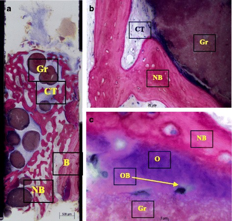
a–c Histological sections of bone core biopsy taken from the site of implantation using a trephine bur. a Overview image of coronal-apical cut through the entire core biopsy showing formation of new bone (NB) next to old bone of the extraction socket (B). easy-graft CRYSTAL particles (Gr) are embedded in well perfused connective tissue (CT) and new bone (NB) (Azur II and Pararosanilin, original magnification ×50). b Integration of easy-graft CRYSTAL particle (Gr) into newly formed bone (NB) and connective tissue (CT) showing tight contact between graft particle and new bone. c High magnification (×200) images of the interface between graft particle (Gr) and new bone (NB) showing osteoblasts (OB) forming osteoid (O) and formation of new bone (NB) on the surface of easy-graft CRYSTAL particles (Gr) (Azur II and Pararosanilin, original magnification ×200)