Figure 3.
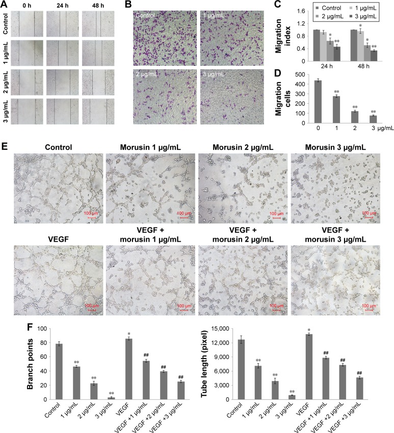
The migration and tube formation inhibition effects of morusin on HUVECs.
Notes: (A, C) Inhibition of HUVEC migration in the wound-healing assay. The photographs were obtained at a magnification of 100×. (B, D) Inhibition of HUVEC migration in the Transwell assay. The penetrated cells were stained and quantified. The photographs were obtained at a magnification of 200×. (E) Reduction in endothelial tube formation after pretreatment with morusin for 24 h following with incubation in 96-well plates with or without VEGF (10 ng/mL). Tube formation of HUVECs was assessed 6–10 h later. The photographs were obtained at a magnification of 200. (F) The histograms represent the dose effect of morusin in branch points and tube length inhibition. *P<0.05 vs control, **P<0.01 vs control. ##P<0.01 vs VEGF-only group. Representative images were taken from three independent experiments.
Abbreviation: HUVECs, human umbilical vein endothelial cells.

