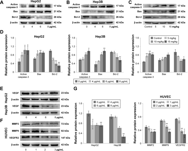
Figure 5.
Apoptotic protein and angiogenic protein expressions in morusin-treated human HCC cells and tumor tissues.
Notes: Western blot analysis of apoptotic protein expression in morusin-treated HepG2 cells (A), Hep3B cells (B), and tumor tissue (C). (D) The histograms show that there was a significant increase in Bax and active caspase-3 and a decrease in Bcl-2 expression in a dose-dependent manner after morusin treatment. (E) Western blot analysis of VEGF expression in HepG2 and Hep3B cells. (F) Western blot analysis of MMP2, MMP, and VEGFR2 expressions in HUVECs. (G) The histograms show that there was a significant decrease in VEGF, MMP2, MMP9, and VEGFR2 expressions in a dose-dependent manner after morusin treatment. *P<0.05, **P<0.01. Three parallel experiments were performed.
Abbreviations: HCC, hepatocellular carcinoma; HUVECs, human umbilical vein endothelial cells.

