
Figure 3.
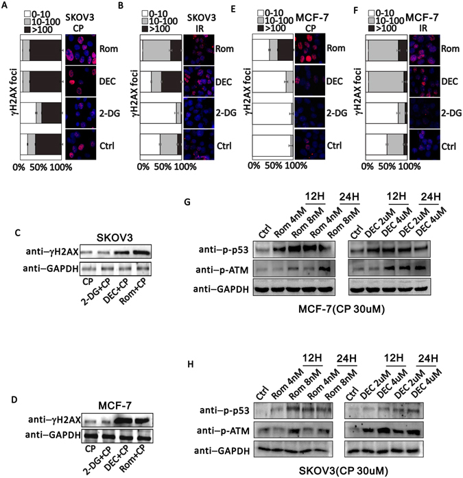
ETD increases DNA damage induced by DTD. (A) γH2AX immunofluorescence staining when cells were exposed to 30 uM CP alone or combining with 2.5 nM Rom, 1 uM Dec or 2 mM 2-DG in SKOV3 cells. (B) γH2AX immunofluorescence staining when SKOV3 cells were exposed to 10 Gy IR alone or combining with 2.5 nM Rom, 1 uM Dec or 2 mM 2-DG. (C and D) Cropped western blots show γH2AX expression level when SKOV3 cells or MCF-7 cells were treated with 30 uM CP alone or combing with 2 mM 2-DG, 1 uM Dec, or 2.5 nM Rom. Uncropped images are in Supplementary information. (E) γH2AX immunofluorescence staining when MCF-7 cells were exposed to 30 uM CP alone or combining with 2.5 nM Rom, 1 uM Dec or 2 mM 2-DG. (F) γH2AX immunofluorescence staining when MCF-7 cells exposed to 10 Gy irradiation alone or combining with 2.5 nM Rom, 1 uM Dec or 2 mM 2-DG. (G and H) Western blotting detection of phosphorylated p53 (p-p53) and phosphorylated ATM upon treatment of MCF-7 (G) and SKOV3 (H) by Rom (left) or DEC (right). GAPDH serves as an endogenous control. 2 time scale (12 H, 12 hours treatment; 24 H, 24 hours treatment) and 2 dosage (Rom, 4 nM and 8 nM; DEC, 2 uM and 4 uM) were applied.