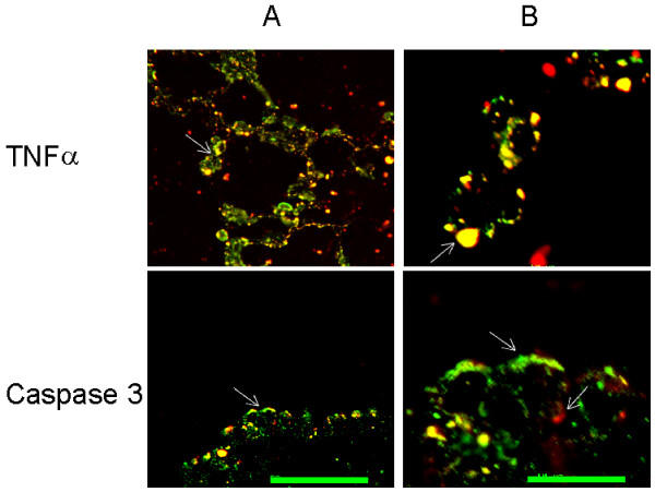
Figure 4.

TNFα is localised in lamellar bodies and caspase 3 in the cytosol of TIIcells. After hyperoxia, rat lungs were fixed and the sections were immunohistochemically double labelled as described in Materials and Methods. A: bar 50 μm; B: higher magnification of the indicated area of A (arrow); bar 10 μm. By confocal microscopy, TIIcells were identified by the green labelling of lamellar bodies (see Methods). The red labelled TNFα appears yellow (arrow in B) when co-localised in lamellar bodies as shown in two TIIcells in B.
