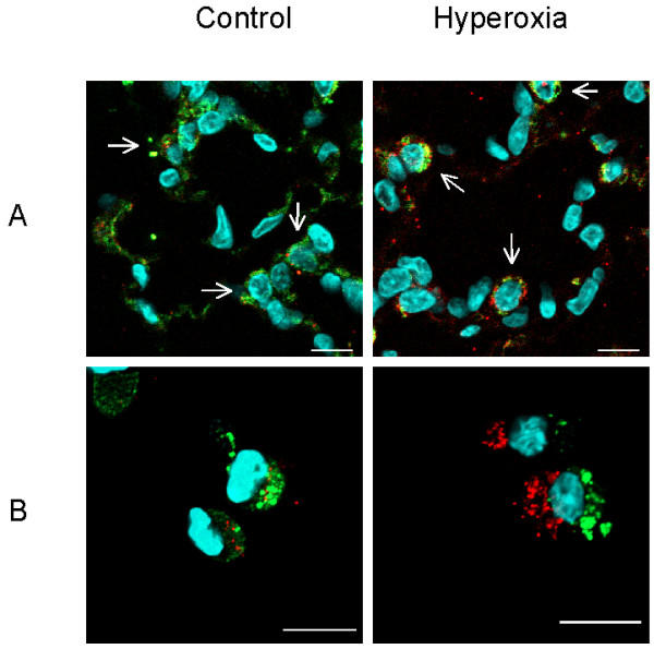
Figure 6.

Hyperoxia activates caspase 3 in TIIcells. Rats were kept normoxic (control) or subjected to hyperoxia. Lung sections (A) and freshly isolated TIIcells (B) were immunohistochemically threefold stained as described in Materials and Methods. Cell nuclei stained light blue (DAPI). TIIcells are distinguishable by the close proximity of their nuclei to green stained lamellar bodies (A; arrows). Active caspase 3 appears red labelled and is predominantly found in the cytosol of TIIcells upon hyperoxic treatment of rats (A and B). Bar 10 μm;
