Figure 1.

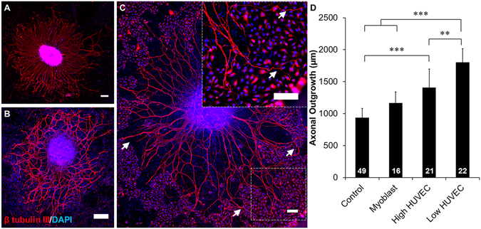
HUVEC enhanced DRG axonal growth is cell type specific. DRG explants cultured on (A) poly-D-lysine (PDL) coated wells, (B) myoblasts, and (C) HUVECs. PDL and myoblast coatings facilitated axonal growth that was radially symmetrical while HUVEC co-cultures facilitated asymmetric growth towards clusters of HUVECs (arrows). Scale = 200 µm. (D) Quantification of axonal growth from DRG explants on different surfaces. Low density of HUVECs supported the most axonal outgrowth of all treatment groups. Data are presented as mean ± standard error. *(p < 0.05), **(p < 0.01), ***(p < 0.001) and brackets indicate significance from other surfaces as determined by one-way ANOVA with Holm-Sidak post hoc analysis (DRG sample size for each group shown in bars from 3 independent replicates).
