
Figure 2.
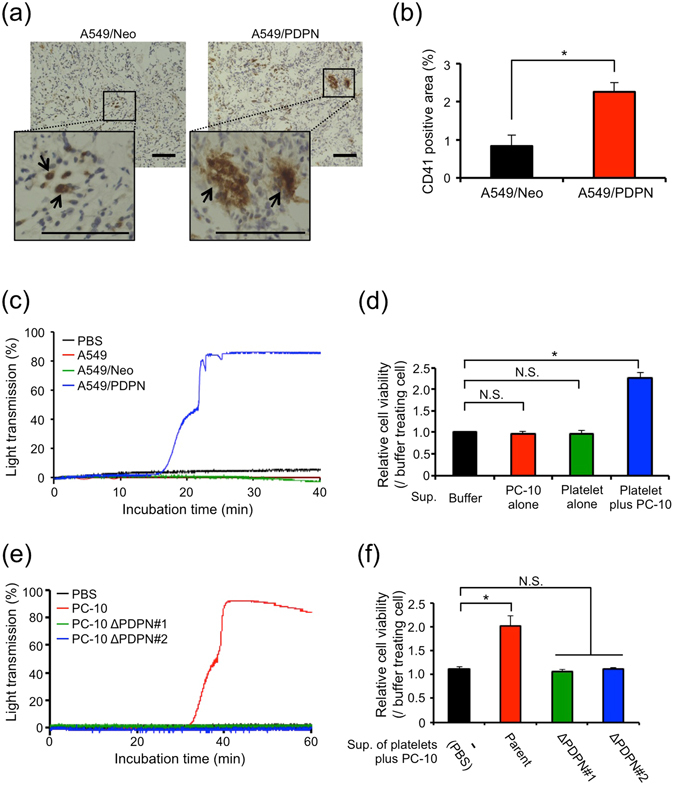
Podoplanin promotes cell growth by enhancing soluble factor secretion from platelets. (a,b) A549 cells that had been transfected with pcDNA3-mock (A549/Neo) or pcDNA3-podoplanin (A549/PDPN) plasmid (5 × 10⁶ cells) were subcutaneously injected into nude mice (N = 3). After 14 days of tumour inoculation, the tumours were extracted and stained with antibodies to CD41 (a). Arrows indicate platelet aggregates. Scale bar: 100 μm. The CD41-positive area was quantitated from 3 independent fields for each mouse (b). All data are shown as means ± SEM. (N = 3) *P < 0.05 by Mann–Whitney U-test. (c) A549/Neo or A549/PDPN cells (2 × 10⁶ cells/ml) were incubated with washed platelets (5 × 10⁸/ml) in Tyrode’s buffer containing 2% murine platelet-poor plasma and 250 μM CaCl2. The light transmission was measured by MCM HEMA TRACER 313 M to monitor the platelet aggregation rate. (d) Supernatants were collected from buffer alone, platelets alone, PC-10 alone, or platelets incubated with PC-10 cells. Then, the PC-10 cells that had been transfected with ZsGreen gene (PC-10/ZsG) were cultured for 72 hours in each supernatant under 0.5% FBS condition. The cell viability of the PC-10/ZsG was calculated from ZsGreen fluorescence. All data are shown as means ± SD of triplicate experiments. *P < 0.05 by Mann–Whitney U-test. N.S.: Not significant. (e) PC-10 and podoplanin-knockout PC-10 (PC-10 ΔPDPN#1 and PC-10 ΔPDPN#2) cells (1 × 10⁶ cells/ml) were incubated with washed platelets (5 × 10⁸/ml) in Tyrode’s buffer containing 2% platelet-poor plasma and 250 μM CaCl2. The light transmission was measured by MCM HEMA TRACER 313 M to monitor platelet aggregation rate. (f) Supernatants were collected from platelets incubated with PC-10 (parent), PC-10 ΔPDPN#1 or PC-10 ΔPDPN#2 cells. Then, PC-10/ZsG cells were cultured with the collected supernatants under 0.5% FBS condition. After 72 hours, the cell viability of the PC-10/ZsG was calculated from ZsGreen fluorescence. All data are shown as means ± SD of triplicate experiments. *P < 0.05 by Mann–Whitney U-test. N.S.: Not significant.