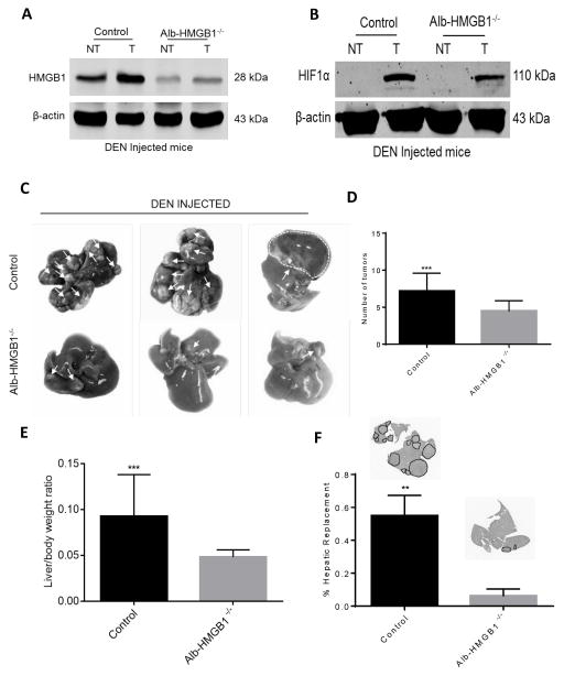
Figure 4. HMGB1 loss in hepatocytes causes decreased tumor growth in mice in response to Diethylnitrosamine (DEN).
(A) HMGB1 levels were significantly increased in tumor tissue compared to liver background in HMGB1 control (HMGB1loxP/loxP) mice injected with DEN 6 months prior. (NT, nontumor liver; T, Tumor). HMGB1 expression is nearly absent in tumor and non-tumor liver tissue in hepatocyte depleted HMGB1 knockout mice (Alb-HMGB1−/−). (B) HIF1α levels are increased in the tumors of both HMGB1 control and Alb-HMGB1−/− mice. (C) Representative images of hepatic nodules (white arrows) after 6 months of DEN treatment in Alb-HMGB1−/− and control mice (D) Alb-HMGB1−/− mice treated with DEN had significantly smaller and less numerous surface nodules compared with HMGB1 control mice (mean 4.5±0.3 nodules in Alb-HMGB1−/− versus 7.2±0.6 nodules in control HMGB1 mice; p<0.001). Alb-HMGB1−/− mice had a significant decrease in tumor burden compared to HMGB1 control mice as seen by (E) liver-to-body ratio (48% decrease in Alb-HMGB1−/− mice, p<0.001) and (F) percentage hepatic replacement by DEN-induced HCC tumors (mean percentage replacement 6.3±1.8% in control vs 55±5.5% in Alb-HMGB1−/−, p<0.01; area occupied by tumors represented as black dashed line). Data represent mean±SEM; n=16 mice/group. The above data are each representative of three experiments with similar results. NS: not significant, **P<0.01, ***P<0.001.

