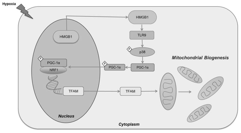
Figure 7. Schematic diagram illustrating the proposed role of HMGB1 and TLR9 in activating mitochondrial biogenesis in hypoxic cancer cells.
During hypoxia, HMGB1 translocated from the nucleus to the cytoplasm of HCC cells. In the cytoplasm, HMGB1 binds and activated TLR9 receptor. This consequently activates TLR9 associated MAP kinases including p38. This leads to the phosphorylation and activation of PGC1-α. Activated PGC1-α then translocates to the nucleus, binds to NRF and both act and transcription co-activators of TFAM. TFAM then translocated to the mitochondria to initiate the process of mitochondrial biogenesis.

