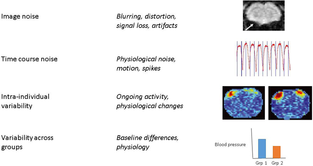
Figure 1. Types of noise.
Image noise refers inaccuracies in the image due to acquisition strategies, including loss of contrast, distortion, and artifacts. The arrow in the image at top indicates distortion in a coronal gradient-echo EPI image of a rat brain acquired at 9.4 T due to the susceptibility mismatch at the air-tissue interface near the ear canals. Time course noise refers to noise that affects images differently at different time points, giving it a temporal structure. Common sources are respiration, cardiac pulsation, and motion. An example respiratory time course is shown. Intra-individual variability may result from changes in baseline physiology, ongoing activity in the brain, or other factors that can change the state of an individual over time. Two representative activation maps from a single rat acquired at different time points in one scanning session are shown at right (adapted from (Matthew Evan Magnuson et al., 2014)). Considerable variability can be seen in experimentally-identical acquisitions. Variability across groups is an important consideration for studies comparing functional neuroimaging data across groups of animals and can result from baseline differences, particularly in factors that affect vascular tone or the relative contribution of physiological noise. A hypothetical difference in baseline blood pressure across two groups is shown for illustration purposes.

