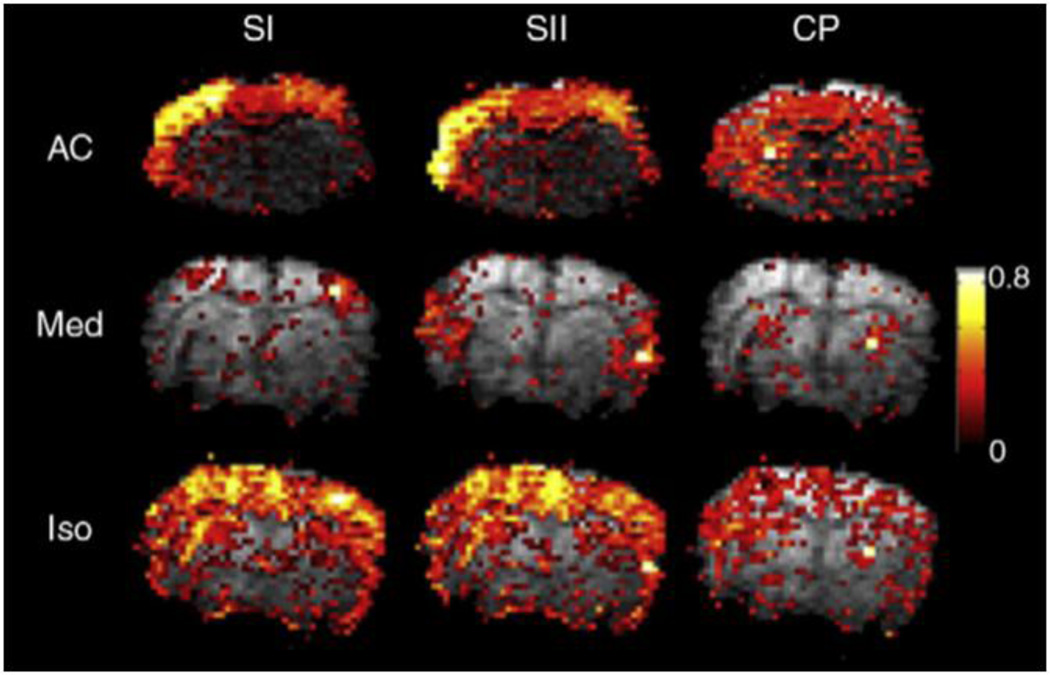
Figure 4. Differences in functional connectivity under different anesthetics.
Connectivity based on seed regions in primary somatosensory cortex, secondary somatosensory cortex, and the caudate putamen are shown for individual rats under three anesthetics (Williams et al., 2010). The strength of the correlation with the seed, the spatial extent, and the specificity vary by anesthetic and by network. Similar effects were observed during group analysis.

