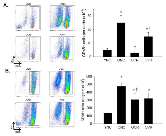
Figure 1. Leukocyte infiltration of aorta and mesenteric vascular arcade.
Aortas (A) and mesenteric vascular arcade (B) from young normal chow (YNC), old- (O) normal chow (NC), voluntary running (VR) and calorie restricted (CR) mice were digested to a single cell suspension and stained with antibodies against CD45 to assess total leukocytes. Representative flow cytometry plots are shown on the left of each panel, summary data is shown on the right. n = 5–11/group. Differences were assessed with one-way ANOVA with LSD post hoc tests. * different from YNC, † different from ONC, p≤0.05. Data are means ± SEM.

