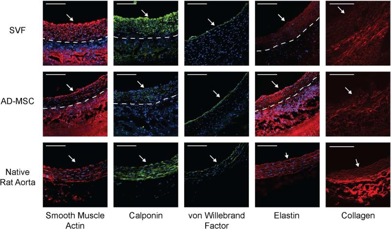
Figure 2. Human SVF-based TEVGs developed into a vascular-like tissue containing the primary cellular and matrix constituents of a small diameter artery, similar to that observed with AD-MSC-based TEVGs.
TEVGs, using either SVF or donor-matched AD-MSCs displayed significant remodeling. TEVGs stained positively for SMCs (smooth muscle actin and calponin), endothelial cells (von Willebrand Factor), and elastin. Multiphoton imaging demonstrated the presence of collagen. White arrow indicates lumen. The dashed line represents the boundary between newly-developed vascular-like tissue (neotissue) and the PEUU scaffold. For comparison, native artery images are included from13.

