
Figure 12.
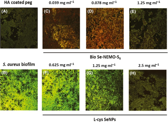
Confocal Laser Scanning Microscopy (CLSM) of HA‐coated peg (A), Staphylococcus aureus biofilm grown onto HA‐coated peg (B) and in the presence of bio6 SeNPs at the concentration of 0.039 mg ml−1 (C), 0.078 mg ml−1 (MBBC) (D), 1.25 mg ml−1 (E), or in the presence of L‐cys SeNPs at the concentration of 0.625 mg ml−1 (F), 1.25 mg ml−1 (G), 2.5 mg ml−1 (H).