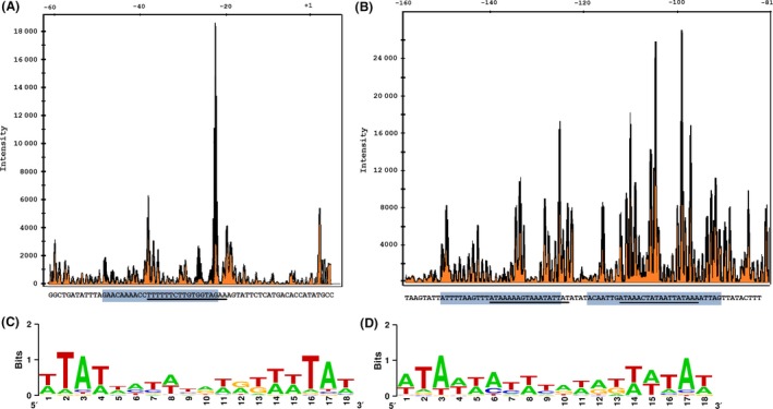
Figure 6.

Identification of the LdtR binding site in (A) PCLIBASIA _04015 and (B) PCLIBASIA _01670 promoters. The DNAse I footprinting electropherograms show a fragment of the digested probe in the absence (orange) or presence (black) of LdtR, highlighting the protected region. The LdtR binding box in each promoter is indicated with a light blue box. Underlined is the palindromic sequence identified with RegPredict. The graphical representation (LOGO) of the linear alignment of LdtR binding sites among the homologues in L. asiaticus of the (C) up‐regulated or (D) down‐regulated genes identified in the RNA‐seq experiments.
