Figure 6.
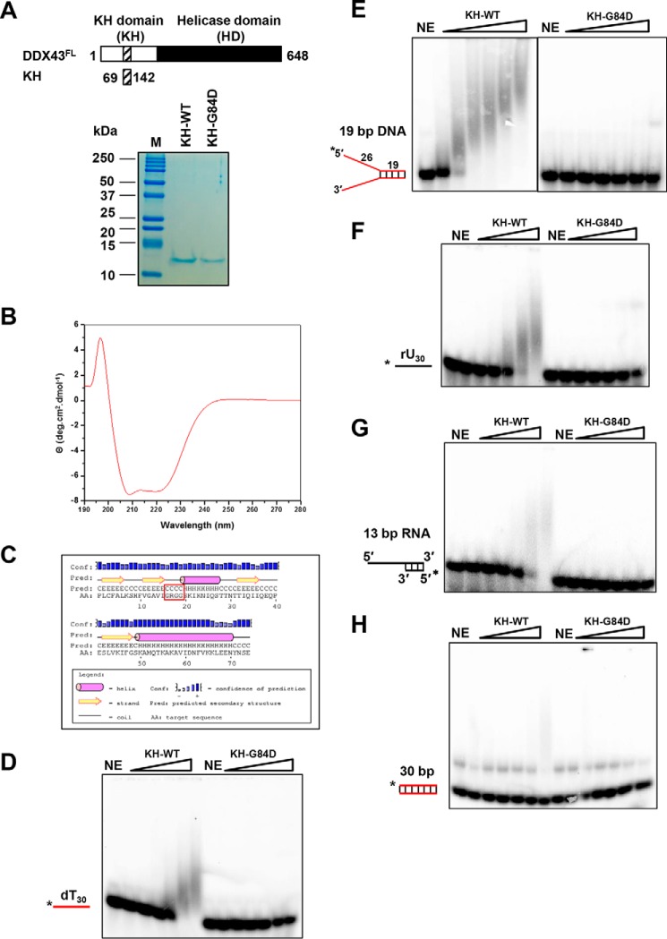
Purification and characterization of DDX43 KH domain. A, schematic representation of full-length DDX43 and its KH domain (top) and purified KH domain proteins (wild type and mutant, 1 μg of protein each loaded) shown on Coomassie-stained SDS-PAGE gel (bottom). NE, no enzyme. B, circular dichroism spectrum of KH domain protein. C, secondary structure prediction of KH domain protein. The motif GRGG is highlighted in red square. D–H, representative EMSA images for KH domain proteins binding with 0.5 nm dT30 ssDNA (D), 19-bp forked duplex DNA (E, two images merged), ssRNA rU30 (F), 5′-tailed 13 bp dsRNA (G), and blunt-end dsDNA (H).

