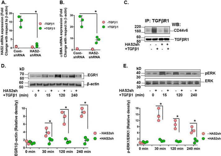
Figure 11.
HAS2-derived HA is required for interaction of CD44V6 with TGFβRI and consequent ERK/EGR1 intracellular signaling. Serum-starved confluent HNLFbs were transfected with control shRNA or HAS2 shRNA for 48 h before treatment with 2.5 ng/ml TGFβ1 for up to 48 h (every 12 h, fresh TGFβ1 was replaced in the medium). A, real-time PCR analysis validates knockdown of HAS2 mRNA. B, real-time PCR analysis demonstrates that HAS2 knockdown inhibits α-SMA mRNA expression. A and B, data are expressed as means ± S.E. (error bars) (n = 3). *, p < 0.05 compared with baseline for each group using Student's two-tailed t test. C, cell lysates were immunoprecipitated (IP) with TGFβRI antibody, followed by WB analysis of CD44v6 and TGFβRI expressions. The blot shown here is representative of three separate experiments. D and E, immunoblotting for EGR1, β-tubulin, pERK, and ERK at the indicated times; the corresponding densitometry graphs are shown in the bottom panels. The blot shown here is representative of three separate experiments, and densitometry graphs show mean ± S.E. of three separate experiments. *, p < 0.01.

