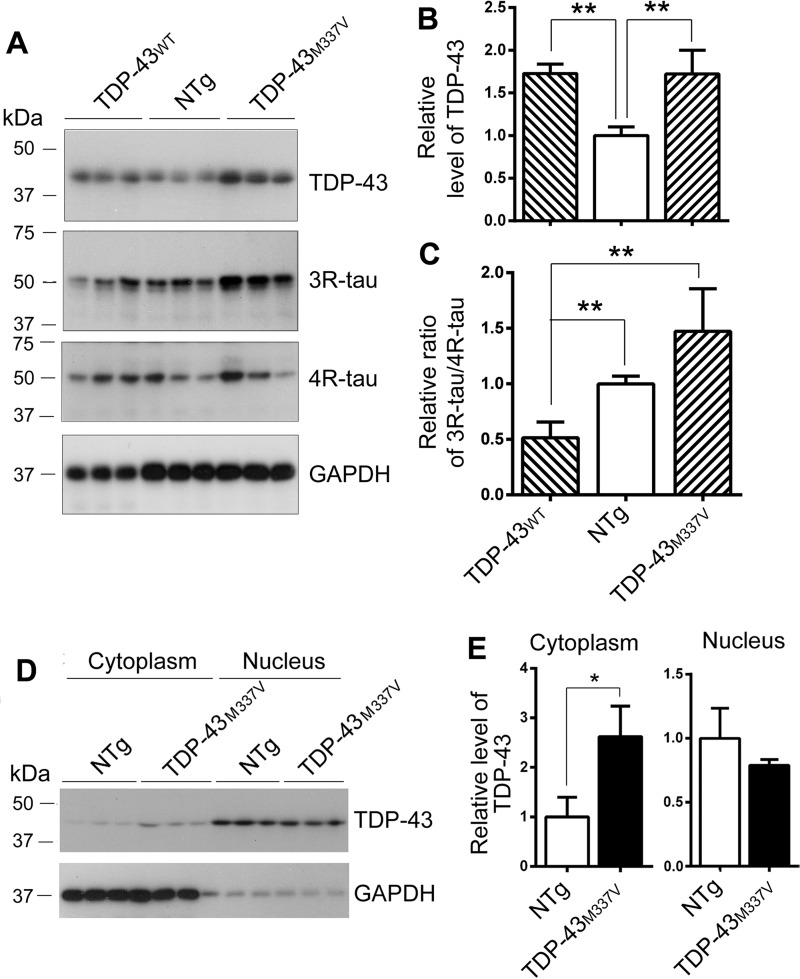
Figure 5.
The ratio of 3R-tau and 4R-tau is decreased in TDP-43 and increased in TDP-43M337V transgenic mouse brains. A–C, the levels of TDP-43 and tau in the brain homogenates from hemizygous human TDP-43WT and mutated human TDP-43M337V transgenic mice and the NTg mice were analyzed by Western blotting. Blots were developed with anti-TDP-43 (A260), anti-3R-tau (RD3), anti-4R-tau (RD4), and anti-GAPDH (A). The relative level of TDP-43 was calculated after normalization with GAPDH (B). The relative ratio of 3R-tau and 4R-tau was calculated after normalization with the corresponding loading controls (C). D and E, the brains from TDP-43M337V transgenic mice and NTg mice were homogenized. The cytoplasm and nucleus were separated by centrifugation. TDP-43 and GAPDH were analyzed by Western blotting (D). The levels of TDP-43 in the cytoplasmic and nuclear fractions were normalized with GAPDH (E). The data are represented as mean ± S.D. (error bars). *, p < 0.05; **, p < 0.01.

