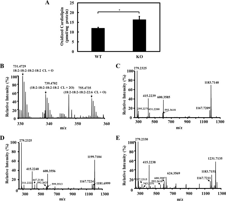
Figure 4.
Genetic ablation of iPLA2γ caused the accumulation of oxidized cardiolipin. A, oxidized cardiolipin levels (i.e. the sum of the three predominant oxidized CL species) in wild-type and iPLA2γ−/− myocardium tissue. Freshly isolated heart tissues from wild-type and iPLA2γ−/− mice were flash-frozen in liquid nitrogen, homogenized using a Teflon pestle grinder, and extracted in the presence of TMCL internal standard. The extracts were purified by aminopropyl solid phase extraction column and analyzed by LC-MS/MS in negative ion mode as described under “Experimental procedures.” Values are the average of four independent preparations ± S.E. *, p < 0.05. B, mass spectrum of oxidized cardiolipin from wild-type mouse myocardium tissue. C–E, aminopropyl solid phase extraction purified lipid extract (from two mouse hearts) was separated on a C18 HPLC column, and the fraction containing oxidized cardiolipin was collected and dried. The dried residue was reconstituted in 50 μl of methanol and analyzed by LC-MS/MS. Fragmentations were performed in the LTQ ion trap with collision energy of 30 eV, and the resultant fragment ions were detected in Orbitrap with a mass resolution of 30,000 at m/z = 400 and a mass accuracy within 5 ppm. MS2 spectra of parent ion [M-2H+]2− at m/z 731 (corresponding 18:2–18:2–18:2–18:2-CL-OH) (C), parent ion [M-2H+]2− at m/z 739 (corresponding 18:2–18:2–18:2–18:2-CL-OOH) (D), and parent ion [M-2H+]2− at m/z 755 (corresponding 18:2–18:2–18:2–22:6-CL-OH) (E) are shown here.

