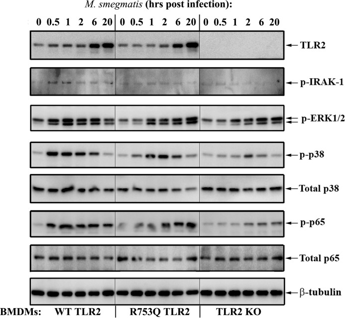
Figure 3.
Macrophages harboring R753Q TLR2 show impaired activation of IRAK-1, MAPKs, and NF-κB upon mycobacterial infection. BMDMs from WT, TLR2 KO, or R753Q TLR2 KI mice were infected for 2 h with M. smegmatis (m.o.i. 10), washed, incubated in gentamycin-containing medium, and lysed at the indicated times after infection. Whole-cell lysates were examined by Western blot analyses with the indicated Abs. The results of a representative experiment (n = 5) are shown.

