
Fig. 6.
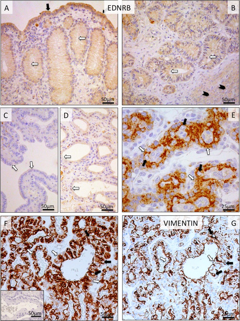
Immunohistochemical (IHC) localization of EDNRB in the canine uterine and utero-placental (Ut-Pl) compartments at selected time points during pregnancy; at the pre-implantation stage (A and B), in the Ut-Pl units during mid-gestation (C, D and E), and in the Ut-Pl compartments at prepartum luteolysis (F). (A, B) At pre-implantation, staining of EDNRB is predominantly localized to the endometrial luminal (surface) epithelial cells (solid arrows in A), glandular epithelial cells of the superficial and deep uterine glands (open arrows in A and B) and myocytes (solid arrowheads in B). During mid-gestation, within the Ut-Pl units, weak endometrial EDNRB expression is detected in the superficial glands (the so-called glandular chambers) and deep uterine glands (open arrows in C and D, respectively). In the placental labyrinth during mid-gestation, strong signals are localized in fetal syncytiotrophoblast cells (open arrows in E). At prepartum luteolysis, only fetal trophoblast (i.e., syncytio- and cytotrophoblast) stains strongly (open arrows in F). No, or sporadically only weak signals, can be identified in maternal decidual cells (solid arrows in E and F). In order to distinguish between fetal and maternal cell types within the canine placenta, i.e., endothelial, trophoblast and decidual cells, VIMENTIN staining was performed on consecutive sections following those used for EDNRB. Endothelial cells (open arrows in G) and decidual cells (solid arrows in G) stain positively for VIMENTIN. There is no background staining in the isotype control (inserted in F).