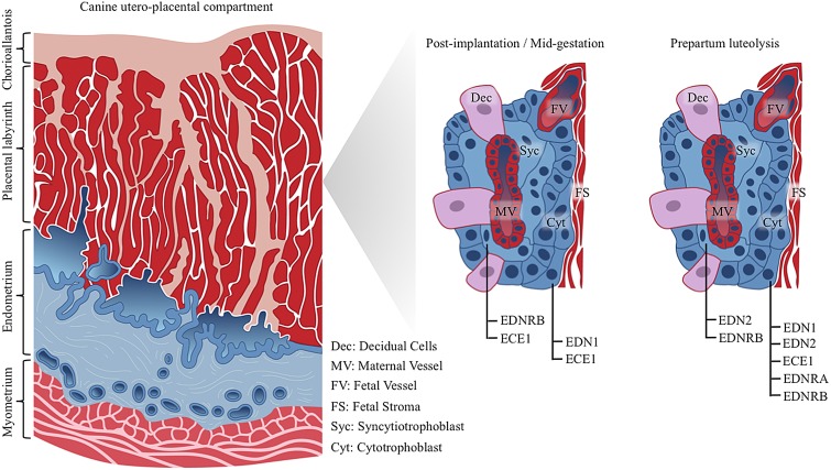
Fig. 9.
Schematic representation of placental EDN system distribution within canine utero-placental compartments (placenta endotheliochorialis). Post-implantation and at mid-gestation similar localization patterns are observed for EDNRB, ECE1 and EDN1. Whereas ECE1 is localized both, in syncytiotrophoblast and cytotrophoblast cells, EDN1 targets only to cytotrophoblast cells, and EDNRB stains positively in syncytiotrophoblast. During prepartum luteolysis, both types of fetal trophoblast cells stain positively for EDN2 and EDNRB. EDN1, ECE1 and EDNRA are localized only in cytotrophoblast cells. As determined by in situ hybridization (ISH), localization patterns of EDN1, ECE1, EDNRA and EDNRB mRNA were similar with expression profiles of the respective proteins (investigated by immunohistochemistry). Due to the lack of commercially available canine-specific antibody, localization of EDN2 was determined at the transcript level by ISH.

