
Fig. 5.
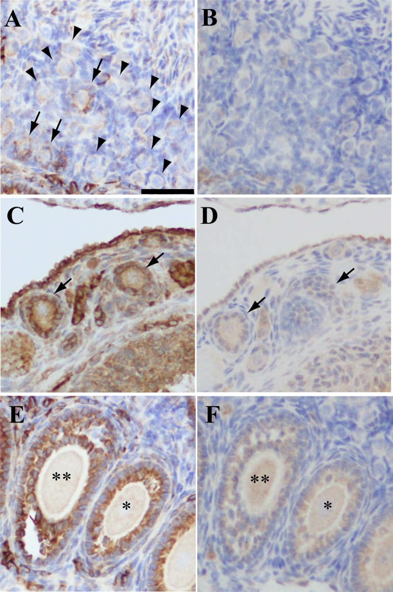
Immunohistochemistry of PGRMC1 in mouse ovary. Panels A and B, C and D, and E and F are images of adjacent mouse ovary sections. Panels A, C and D: stained with anti-PGRMC1 antibody. Panels B, D and F: stained with rabbit IgG. Panel A: arrows, primordial follicles expressing PGRMC1 in granulosa cells; arrowheads, primordial follicles without PGRMC1 expression. Panel C: arrows, primary follicles expressing PGRMC1 in granulosa cells; Panel D: arrows, same follicles indicated in panel C. Panel E and F: * secondary follicle; ** antral follicle expressing PGRMC1 in granulosa cells. Scale bar: 50 μm.