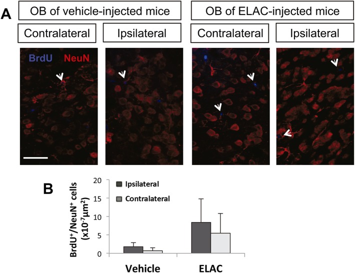
Figure 9.

Effect of in vivo administration of ELAC on newly formed OB neurons. Adult mice were injected in the right lateral ventricles with ELAC (5 μM, 2 μL; n = 6) or vehicle (n = 6) and then injected with the cell‐division marker BrdU (120 mg·kg−1) during 3 days after treatment administration starting on the day of the surgical procedure and were killed on day 10 after surgery. (A) Fluorescence microscopy images of brain coronal sections showing the ipsilateral and contralateral OBs of mice treated with ELAC or vehicle; sections were processed for immunohistochemical detection of BrdU+ nuclei (red) and NeuN (blue). (B) Quantification of BrdU+/NeuN+ cells within the SVZ of mice that had received i.c.v. injections of ELAC or vehicle.
