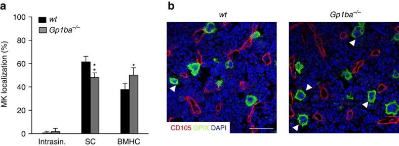
Figure 1. GPIbα deficiency alters MK localization in the BM.
(a) Quantification of MK localization in the BM reveals less sinusoidal contact (SC) and increased localization in the BM haematopoietic compartment (BMHC) in Gp1ba−/− mice (grey) compared to the wild-type (wt, black) (n=5); intrasinusoidal (intrasin.). (b) Representative confocal images of immunostained BM of wt and Gp1ba−/− mice. Scale bars, 50 μm. MKs, proplatelets and platelets are shown by CD41 staining in green. Endoglin staining (red) labels vessels. DAPI, blue. Arrowhead indicates MKs in the BM haematopoietic compartment (BMHC). Bar graphs represent mean±s.d. Two-way ANOVA with Bonferroni correction for multiple comparisons; **P<0.01; *P<0.05.

