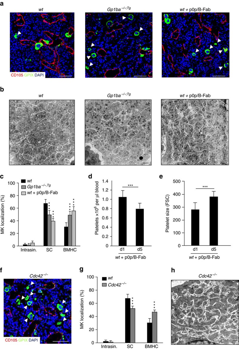
Figure 2. GPIbα ectodomain and Cdc42 regulate MK localization in the BM.
(a) Confocal images of immunostained BM and (b) TEM analysis of BM MKs of wt (left panel), Gp1ba−/−;tg (Gp1ba-Tg) (middle panel) and wt mice after treatment with GPIbα-blocking monovalent Fab fragments, p0p/B-Fab (right panel). n=4 biological replicates. Scale bars, 50 μm (a) and 2 μm (b). MKs, proplatelets and platelets are shown by GPIX staining in green. Endoglin staining (red) labels vessels. DAPI, blue. Arrowhead indicates MKs in the BM haematopoietic compartment (BMHC). (c) Quantification of MK localization in the BM reveals less sinusoidal contact (SC) in Gp1ba-Tg (dark grey) and wt mice after GPIbα-blockade (light grey) compared to the wt (black) (n=7, 5 and 10); intrasinusoidal (intrasin.). (d,e) Reduced platelet count (d) and increased platelet size (e) in wt mice after GPIbα-blockade (n=4). (f) Representative confocal images of immunostained BM of Cdc42−/− mice. Scale bar, 50 μm. (g) Quantification of MK localization in the BM reveals reduced SC in wt (black) and Cdc42−/− (grey) mice (n=10 and 3). (h) TEM analysis of BM MKs of Cdc42−/− mice. Scale bar, 2 μm. Bar graphs represent mean±s.d. (c,g) Two-way ANOVA with Bonferroni correction for multiple comparisons; (d,e) Unpaired two-tailed Student’s t-test; ***P<0.001.

