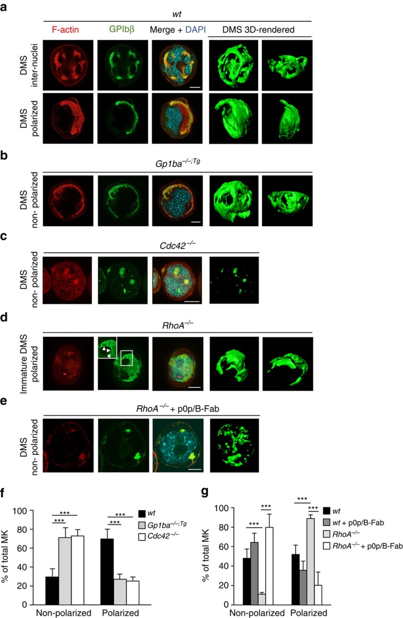
Figure 5. RhoA controls GPIbα/Cdc42-dependent MK polarization.
(a–e) Representative confocal images of in vitro differentiated MKs reveal polarized DMS in mature wt MKs (a), defective polarization in Gp1ba−/−;Tg (Gp1ba-Tg) (b) and Cdc42−/− MKs (c), increased polarization in RhoA−/− MKs (d) and defective polarization of RhoA−/−MKs after treatment with GPIbα-blocking monovalent Fab fragments, p0p/B-Fab (e). F-actin is stained by phalloidin (red); GPIbβ (a–d) or GPIX (e), green; DAPI, blue. 3D surface rendering of respective z-stacks (Imaris software; right panel). n=5 biological replicates. Scale bar: 10 μm. (f) Quantification of DMS polarization shows reduced MK polarization in Gp1ba-Tg (light grey) and Cdc42−/− (white) MKs compared to the wt (black) (n=5 biological replicates). (g) MK polarization is increased in RhoA−/− MKs (light grey) compared to the untreated wt (black) and wt MKs treated with p0p/B-Fab fragments (dark grey). Treatment of RhoA−/− MKs with p0p/B-Fab fragments (white) reverts the hyperpolarization. n=5 biological replicates (wt and RhoA−/−) and 2 biological replicates (wt+p0p/B-Fab and RhoA−/−+p0p/B-Fab). Bar graphs represent mean±s.d. Two-way ANOVA with Bonferroni correction for multiple comparisons; ***P<0.001.

