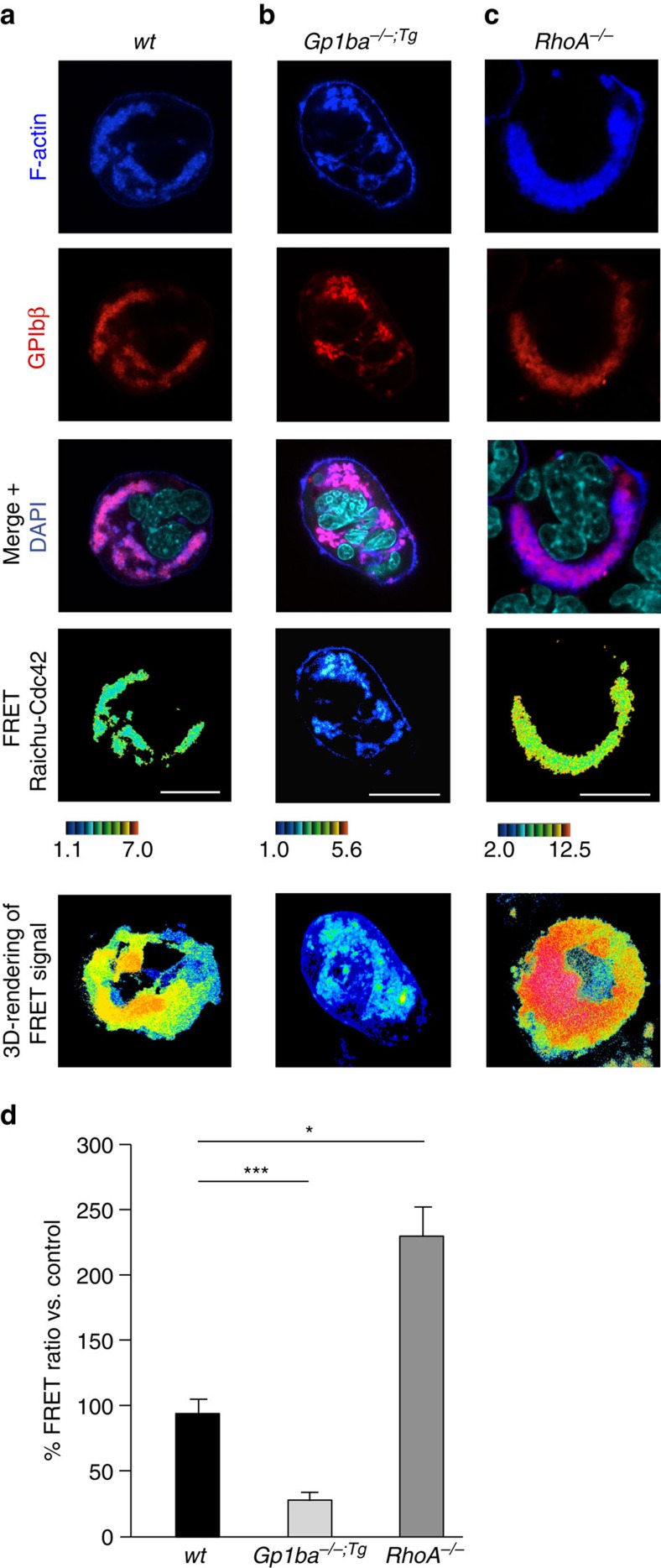
Figure 6. RhoA controls GPIbα-induced MK polarization by limiting Cdc42 activity.
(a–c) Cultured mouse MKs were transduced with Raichu-Cdc42 lentiviral vector. F-actin was stained with phalloidin (blue); GPIbβ, red; DAPI, cyan. Cdc42 activation was visualized by colour-coded FRET. High ratio values (red) correlate with higher Cdc42 activity. Active Cdc42 localizes with the polarized F-actin/DMS complex and its activity is decreased in Gp1ba−/−;Tg (Gp1ba-Tg) and increased in RhoA−/− MKs. Scale bar: 10 μm. (d) Quantification reveals decreased Cdc42 activity in Gp1ba−/−;Tg (light grey) and increased activity in RhoA−/− (dark grey) polarized MKs compared to the wt (black) (n=4 biological replicates). Bar graphs represent mean±s.d. Two-way ANOVA with Bonferroni correction for multiple comparisons; *P<0.05; ***P<0.001.

