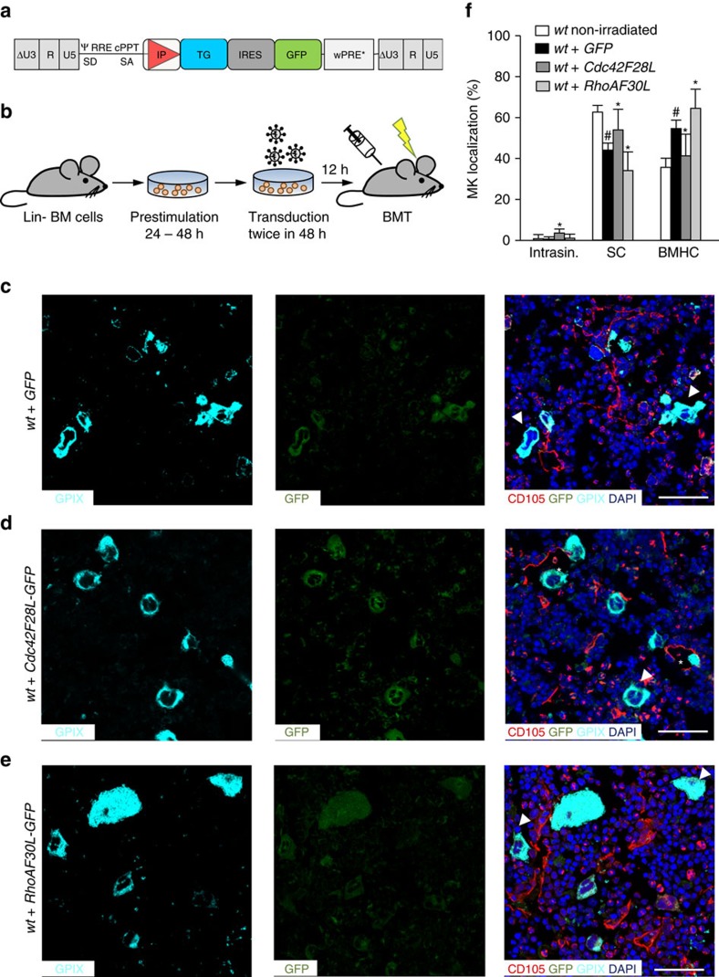
Figure 7. Activation states of RhoA and Cdc42 have opposing functions in MK localization.
(a) Third-generation self-inactivating lentiviral vectors were generated, expressing Cdc42F28L and RhoAF30L, respectively, together with GFP through an IRES, or just GFP under control of the MK-specific human GP6 promoter in the internal position. (b) Work-flow. Lineage marker-negative (Lin−) BM cells were isolated, pre-stimulated for 24 h and transduced twice. Cells were then transplanted into conditioned recipient mice. (c–e) Representative confocal images of immunostained BM of wt mice after BM transplantation of wt HSC transduced with GFP (c), constitutive active Cdc42 (F28L) (d) or constitutive active RhoA (F30L) (e). Scale bars, 50 μm. MKs, proplatelets and platelets are shown by GPIX staining in cyan colour. Endoglin staining (red) labels vessels. DAPI, blue. Arrowhead indicates MKs in BMHC, asterisk indicates intrasinusoidal MKs. (f) Quantification of MK localization in the BM (n=4). Quantification of MK localization in non-irradiated wt mice (white) are shown to demonstrate altered localization after irradiation and transplantation of wt cells. Bar graphs represent mean±s.d. Two-way ANOVA with Bonferroni correction for multiple comparisons. *P<0.05, compared to wt+GFP; #P<0.05, compared to wt not transplanted.

