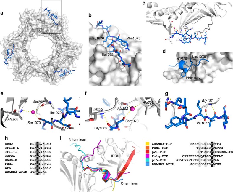
Figure 7. Structure of the PCNA:ZRANB3(APIM) complex.
(a) Front view of the PCNA ring (grey surface and ribbons) with the ZRANB3 APIM motif peptide (blue sticks). (b) Magnified view of the boxed region in e. Shown is a surface representation of one of the three APIM motif binding sites on the PCNA ring with the bound APIM motif peptide (blue sticks coloured by atom type). (c) Overview of the hydrogen-bond interaction network between the ZRANB3 APIM motif peptide (blue) and PCNA (grey). Bound water molecule (purple sphere) and hydrogen bonds (yellow dotted lines) are shown. (d) Hydrophobic pocket on PCNA surface (grey) with the residues that form ‘hydrophobic plug’ in the APIM motif (Ile 1072, Phe1075 and Leu1076; shown as blue sticks). (e–g) Magnified view of the hydrogen-bond interaction network between the ZRANB3 APIM motif peptide (blue) and PCNA (grey, labels in italics). Bound water molecule (purple sphere) and hydrogen bonds (yellow dotted lines) are shown. (h) Alignment of the APIM motifs from different human proteins. (i) Superimposed backbones of bound PIP box and APIM motif peptides from different co-crystal structures. Alignment of the peptides is shown on the right. Shown are ZRANB3-PIP515–529 (yellow; PBD:5MLO), FEN1336–348 (orange; PDB:1U7B), p21143–160 (red; PDB: 1AXC), Polη694–713 (magenta; PDB: 2ZVK), p1551–71 (cyan; PDB: 4D2G) and ZRANB3-APIM1065–1079 (blue; PDB:5MLW).

