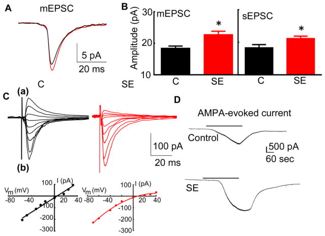
Figure 1. AMPAR-mediated neurotransmission of CA1 pyramidal neurons was enhanced in animals in SE.
SE was induced in adult male rats by a high dose of pilocarpine (350 mg/kg). Brain slices were prepared 40 minutes after the first behavioral seizure and were allowed to recover for 20 min. (A) The averaged action potential-independent synaptic currents (mEPSCs) recorded from CA1 pyramidal neurons of an animal in SE (red) and a naïve animal (black). (B) Mean of the median mEPSC and sEPSC amplitude. The values represent the mean ± SEM (N= 11 neurons from 9 SE animals and 13 neurons from 7 naïve animals for mEPSCs, * p < 0.05, t-test and N= 9 neurons of 7 SE animals and 9 neurons of 6 animals for sEPSCs,* p < 0.05. t-test). (C) Rectification properties of evoked EPSCs recorded from CA1 neurons of control (black) and SE (red) animals. The neurons were held at different voltages (-70 mv to +40 mV) and EPSCs were evoked (eEPSCs) by electrical stimulation of Schaffer collaterals. The eEPSCs recorded from the CA1 neuron of a representative control (black) and SE (red) animal are shown in (a). The amplitude of currents recorded in the same cells displayed in (a) plotted against the holding potentials are shown in (b). (D) Representative whole-cell AMPA-evoked current recorded from CA1 pyramidal neurons of SE (60 min after the first stage 5 behavioral seizure) and naïve animals. AMPA (2.5 μM, black bar) was bath applied after recording a baseline holding current.

