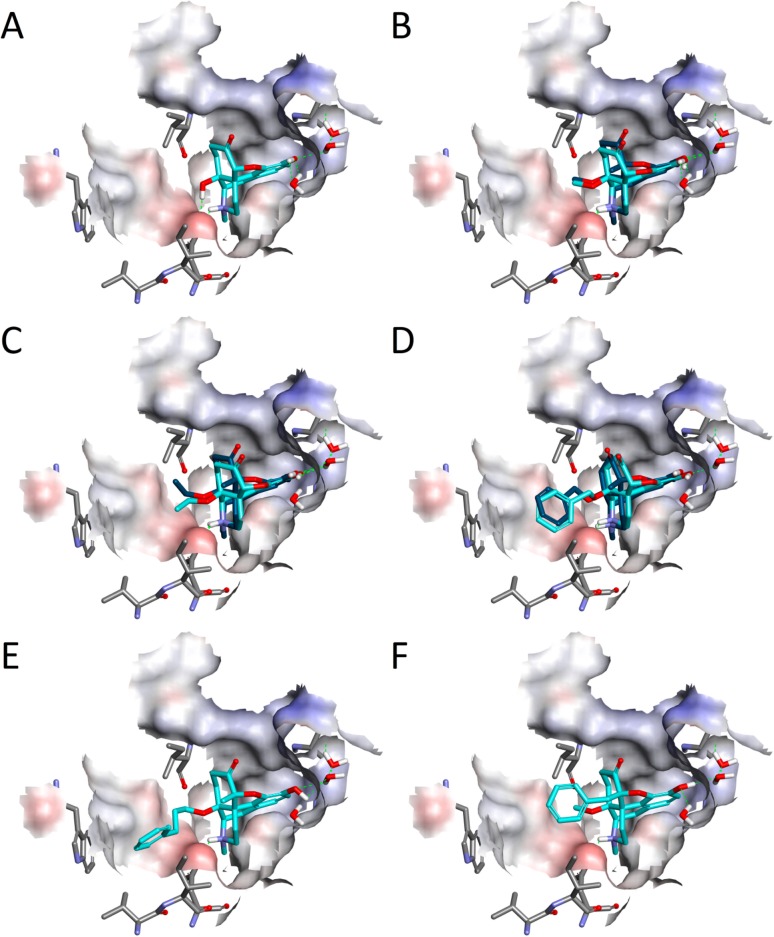
Figure 4.
Docking of the investigated 14-oxygenated N-methylmorphinan-6-ones to the active crystal structure of the μ-OR. Shown are the binding poses of (A) 1 (OM, c), (B) 2a (14-OMO, c) and 2b (14-MM, b), (C) 3a (14-OEO, c) and 3b (14-EM, b), (D) 4a (14-OBO, c) and 4b (14-BM, b), (E) 5 (PPOM, c), and (F) 6 (BOMO, c), where c/b denotes cyan/blue. Hydrogen bonds are depicted as green dashed lines, and the binding pocket surface is color-coded according to the interpolated charge (blue/red = positive/negative).

