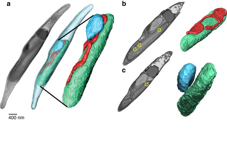
Figure 4. Three-dimensional organization of a P. tricornutum cell.
(a) Whole cell reconstruction of an intact P. tricornutum cell based on FIB-SEM images reveals the physical contacts between the chloroplast (green), mitochondrion (red) and nucleus (blue). (b) Chloroplast–mitochondria interaction. (c) Chloroplast–nucleus interaction. Images represent frames from Supplementary movie 1. Grey pictures in a, stacks of SEM micrographs; in b,c: selected single SEM frame. Coloured pictures in a–c: 3D reconstruction. Yellow arrows highlight contacts between organelles. Bar: 400 nm.

