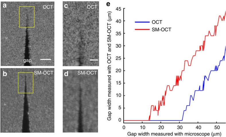
Figure 2. SM-OCT demonstration of improved visibility of closely spaced scattering objects.
(a,b) A phantom composed of PDMS and TiO2 powder was shaped to form a gap of decreasing size to evaluate the effective spatial resolution of SM-OCT versus OCT. The images shown here are en face OCT (a) and SM-OCT (b) scans inside the phantom. Scale bar, 100 μm. (c,d) Close-up view on the regions marked in a,b showing the micron-size gap. Scale bar, 50 μm. The gap that is clearly visible in SM-OCT does not appear in the OCT image due to speckle noise. (e) The size of the gap measured from the OCT and SM-OCT images versus the size of the gap measured from a bright-field microscope image (10 × , NA=0.25). The minimum size of a resolvable gap is decreased by a factor of 2.5 owing to SM-OCT. Note that the visibility of the gap is limited only by the speckle created by the turbid PDMS-TiO2 phantom.

