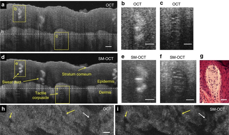
Figure 7. SM-OCT imaging of intact human fingertip skin reveals fine structures such as the tactile corpuscle.
(a) OCT B-scan of a fingertip. Scale bar, 100 μm. (b) Close-up view on the sweat duct marked in a. Scale bar, 50 μm. (c) Close-up view on the tactile corpuscle marked in a. Scale bar, 50 μm. (d) SM-OCT scan of a fingertip. (e) Close-up view on the sweat duct marked in d. Scale bar, 50 μm. (f) Close-up view on the tactile corpuscle marked in d. Scale bar, 50 μm. (g) Microscope image of H&E-stained tactile corpuscle (courtesy of Dr Jesus Lozano, Dr Lorena Monarrez and Professor Doug Schmucker, Department of Anatomy, UCSF School of Medicine). Scale bar, 50 μm. (h,i) OCT and SM-OCT en face images from a 3D scan of the fingertip, located at the top of the dermis, as shown by the dashed line in a,d. With SM-OCT, there is an improved delineation of the dermal papillae (yellow arrows) and sweat ducts (white arrow). Scale bar, 100 μm.

