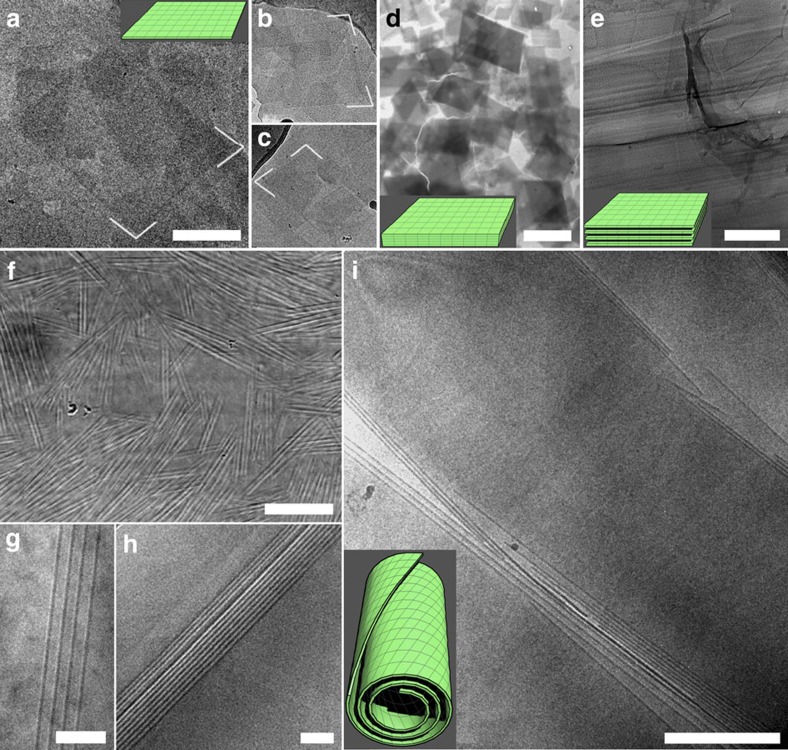
Figure 2. Morphologies of the nanosheet-based and tubular structures formed by SDS@2beta-CD complexes.
(a–c) Cryo-TEM pictures of parallelogram nanosheets with an obtuse angle of 104°, scale bar, 200 nm. (d) A TEM image of flake crystals with a shape identical to that of the nanosheets, scale bar, 5 μm. (e) A freeze-fractured TEM picture of lamellar structures, scale bar, 100 nm. (f–i) Pictures of multilamellar tubular structures: an overview (f, optical microscopy, scale bar, 20 μm), zoom-in of the tube walls (g and h, cryo-TEM, scale bar, 100 nm), and a fractured tube (i, cryo-TEM, scale bar, 500 nm).

