Fig. 2.

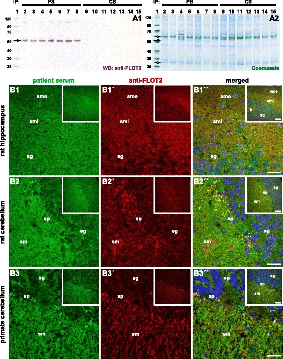
Histo-immunoprecipitation and antigen identification. Cryosections of rat or pig cerebellum were incubated with the serum (1:100), washed in PBS, and solubilized using detergents. The solution was incubated with protein-G-coated magnetic beads. The immunocomplexes were eluted by SDS and subjected to SDS-PAGE analysis and Western blot. A Western blot after incubation with anti-flotillin-2 (A1) and enzymatic visualization of antibody binding. Staining of SDS polyacrylamide gel with colloidal Coomassie (A2). Lane 1: molecular mass (kDa) marker; lanes 2–8: histo-immunoprecipitates of patient sera from rat cerebellum; lanes 9–15: histo-immunoprecipitates of control samples. The arrow indicates the position of the immunoprecipitated antigen 50 kDa while dotted arrows indicate the position of IgG heavy and light chain at 52 and 27 kDa, respectively. PS patient sample, CS control sample. B Immunofluorescence staining of rat hippocampus (B1) and cerebellum (B2) and primate cerebellum (B3) tissue sections with patient serum (green, 1–3) and anti-flotillin-2 antibody (red, 1′–3′). The merged images show localization of the reactivity in the same region including the more intense staining of the sm internum on the hippocampus (1″–3″). Scale bar: 50 μm (large images), 100 μm (inserts). PS patient serum, CS control serum, h hilus, sg stratum granulosum, sm stratum moleculare, smi stratum moleculare internum, sme stratum moleculare externum, sp stratum purkinjense
