Fig. 2.

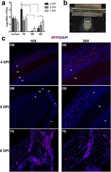
HSV-1 infected cells are observed within the glomerulus of the OB. a Infectious virus was measured by plaque assay from the corneas, TG, OB, and BS at 3, 5, and 7 DPI following infection with 1000 PFU HSV-1/cornea of C57BL/6 mice (corneas, n = 4–7/group, TG, n = 6–9/group, OB n = 6–8/group, BS, n = 8–12/group). **p < 0.005 was determined by using a Dunn’s multiple comparison’s post-test between tissue at the specified time points and ^p < 0.05 comparing 3 and 7 DPI from the same tissue using a Dunn’s multiple comparison post-test following a Kruskal-Wallis one-way analysis of variance. b, c RosaTd/Tm mice (n = 2/time point) were infected with 200 PFU SC16 ICP0-Cre-expressing virus/cornea. b Image illustrates transverse sectioning of whole brain tissue for microscopy analysis (white box depicts OB). c Representative images of RFP+ cells infected with virus localized within the glomerular layer of the OB (Gl) at 4 DPI and 6 DPI. The bottom panels display abundant RFP+ labeling within the TG from longitudinally sectioned tissue at 6 DPI. Nuclei are stained with DAPI. White scale bar indicates 40 μM. Other layers of the OB include the external plexiform layer (Epl), mitral cell layer (Mi), and the granule cell layer (Gr)
