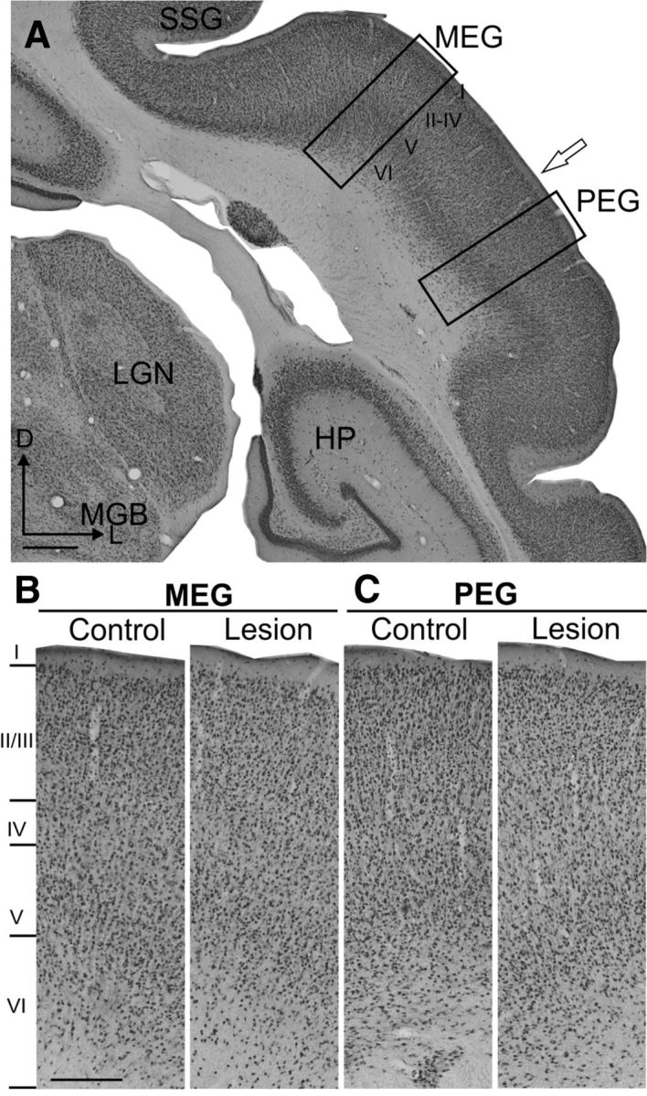
Figure 7.

Neuronal density in layer VI of MEG is lower in animals with corticothalamic lesions than in controls. A, Coronal section immunostained with NeuN at the level of the auditory cortex (control case). Rectangles indicate the regions of MEG (where A1 is located) and PEG shown at higher magnification in B and C. The open arrow indicates the border between MEG and PEG. B, NeuN cell density in MEG layer VI of the lesion example is lower than in the control (for quantification, see Fig. 6). C, No difference in cell density in layer VI of the PEG was found between control and lesion cases. Scale bars: A, 1 mm; B, 250 μm. I–VI, layers 1–6 of the cortex.
