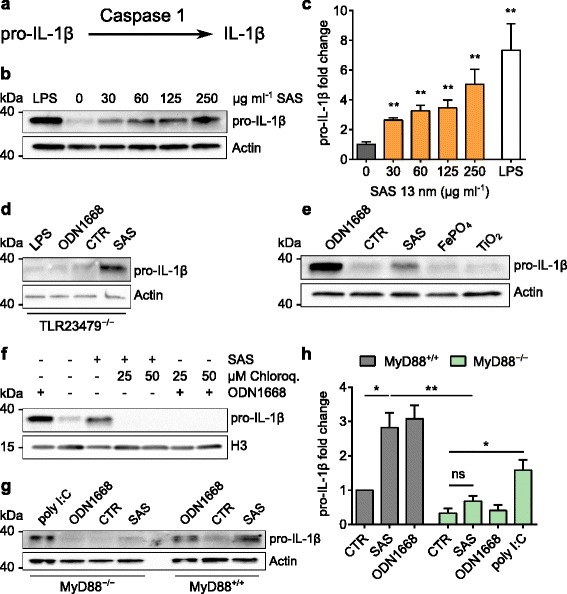
Fig. 4.

Induction of pro-IL-1β by food-grade SAS particles depends on MyD88. Immature DCs were incubated (18 h, 37 °C) with particles to test for IL-1β induction. Asterisks denote significant differences between SAS treatments and controls (*p < 0.05, **p < 0.01, ***p < 0.001, ****p < 0.0001). a Schematic illustrating the mechanism of IL-1β production. b DCs were incubated with LPS (250 ng ml−1) or 13-nm SAS and analyzed for pro-IL-1β (31 kDa) and actin (42 kDa) by immunoblotting. c Quantification of pro-IL-1β induction by SAS particles (unpaired two-tailed t-test, n = 5, error bars, s.e.m.). d Incubation of TLR2/3/4/7/9−/− DCs with LPS (250 ng ml−1), ODN1668 (600 ng ml−1), medium (CTR) or SAS particles (125 μg ml−1). e Incubation of wild type DCs with ODN1668 (600 ng ml−1), medium or the indicated particles (125 μg ml−1). f Effect of endosomal TLR inhibition. DCs were incubated with 13-nm SAS (125 μg ml−1) alone or in the presence of chloroquine and analyzed for pro-IL-1β and histone H3 (17 kDa) by immunoblotting. ODN1668 (600 ng ml−1) served as positive control. g MyD88−/− or wild type (MyD88+/+) DCs were incubated with poly I:C (5 μg ml−1), ODN1668 (600 ng ml−1), medium (CTR) or SAS particles (125 μg ml−1) and analyzed for pro-IL-1β (31 kDa) and actin (42 kDa) by immunoblotting. Split bands in some control lanes are an electrophoretic artifact not interfering with quantifications. h Pro-IL-1β induction by SAS particles in MyD88−/− or wild type (MyD88+/+) DCs (unpaired two-tailed t-test, n = 3, error bars, s.e.m.)
