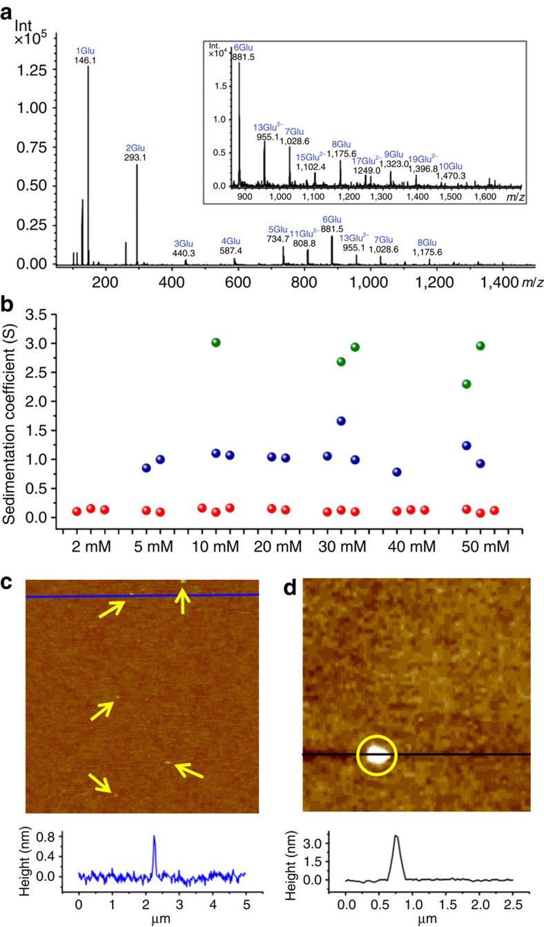
Figure 1. Evidences of different cluster species by using different analytical tools.
(a) ESI Ion-Trap mass spectrum (negative ion mode) of a 40 mM solution of DL-Glu in water at pH 11 (adjusted with NH4OH). Inset: zoom into the high m/z range. (b) Sedimentation coefficients of species detected by AUC at 60,000 r.p.m. in D2O solutions of DL-Glu at different concentrations, as resulting from two or three independent experiments (see Supplementary Fig. 3 for corresponding results in H2O). (c,d) The in situ AFM images of species deposited from a supersaturated solution of DL-Glu on the (100) surface of a silicon wafer. The plots at the bottom are height profiles along the blue and black lines in the images. Fields of view: 5 μm in c and 2.5 μm in d.

