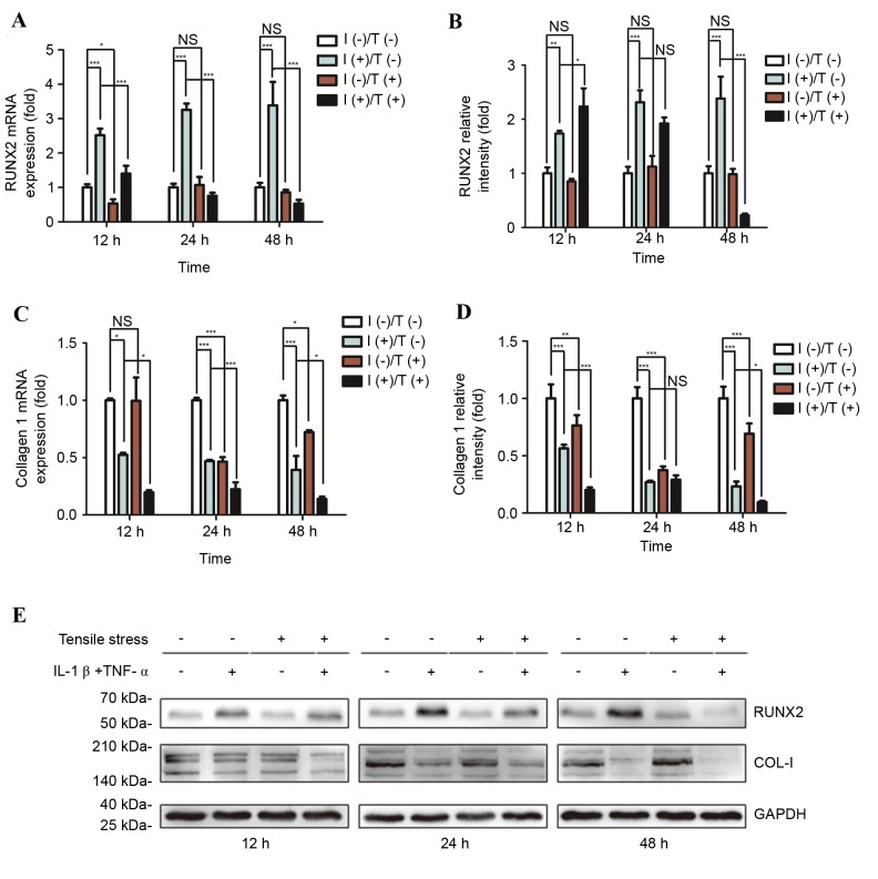
Figure 4.
Differential expression of RUNX2 and COL-I in response to pro-inflammatory stimuli and/or tensile strength. Relative osteogenic and matrix gene expression and protein levels of RUNX2 and COL-I in PDLCs were assessed. (A) mRNA and (B) protein expression levels of RUNX2, and (C) mRNA and (D) protein expression levels of COL-I were determined following treatment for 12, 24 or 48 h. (E) Representative western blot images of RUNX2 and COL-I protein expression levels. GAPDH served as an internal control. Data are expressed as the mean ± standard deviation (n=3). *P<0.05, **P<0.01, ***P<0.001. I(−)/T(−), untreated cells; I(−)/T(+), cells treated with tensile strength alone; I(+)/T(−), cells treated with IL-1β/TNF-α alone; I(+)/T(+), cells treated with tensile strength and IL-1β/TNF-α at each time point. PDLCs, periodontal ligament cells; IL-1β, interleukin-1β; TNF-α, tumor necrosis factor-α; NS, non-significant; RUNX2, runt-related transcription factor 2; COL-I, type I collagen.

