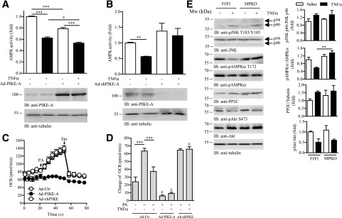
Figure 5.
PIKE-A is involved in TNF-α–mediated AMPK inactivation and lipid oxidation. A: C2C12 myotubes were infected with control adenovirus (Ad-Ctr) or Ad-PIKE-A. Forty-eight hours after infection, the cells were further stimulated with TNF-α (10 ng/mL) for 24 h. The AMPKα were then immunoprecipitated and used for the in vitro kinase assay. Results were expressed as fold change against the PBS-treated Ad-Ctr–infected cells (top panel; *P < 0.05, ***P < 0.001, one-way ANOVA; n = 3). Expression of PIKE-A and tubulin was also determined by immunoblotting (IB; bottom panel). B: C2C12 myotubes were infected with Ad-Ctr or Ad-shPIKE. Forty-eight hours after infection, the cells were further stimulated with TNF-α (10 ng/mL) for 24 h. The AMPKα were then immunoprecipitated and used for the in vitro kinase assay. Results were expressed as fold change against the PBS-treated Ad-Ctr–infected cells (top panel; **P < 0.01, one-way ANOVA; n = 3). Expression of PIKE-A and tubule was determined by Western blot analysis (bottom panel). C: A representative profile of OCR in cells infected with Ad-Ctr, Ad-PIKE-A, or Ad-shPIKE for 48 h. The times when PA (1 mmol/L) and etomoxir (Eto; 40 μmol/L) were injected are indicated by arrows (n = 4). D: Changes of OCR in Ad-Ctr–, Ad-PIKE-A–, or Ad-shPIKE–infected C2C12 myotubes after stimulation by various combinations of TNF-α (10 ng/mL, 24 h) and PA (1 mmol/L) (***P < 0.01; cP < 0.001 vs. Ad-Ctr–infected cells of the same treatment, Student t test; n = 4–6). E: Immunoblotting analysis of liver and hind limb muscle (mixed fiber type) collected from Fl/Fl and MPKO mice after PBS or TNF-α (1 µg/kg/day) infusion for 24 h. Expression and phosphorylations (p) of JNK, AMPK, PP2C, Akt, and tubulin were also verified. Quantification of the Western blot analysis is shown in the right panel (**P < 0.01, one-way ANOVA, n = 3).

